North Carolina State University
![]()
| PhD. Chemistry,
University of North Carolina |
 |

Michigan State University

Amherst College

| B.S. Chemistry,
North Carolina State University
|
|
|
post-doctoral research,
Michigan State University
|
post-doctoral research,
Amherst College
|
|
|
Bioinorganic Chemistry: A Personal Journey |
|
|
"Only God knows how to use the First-row transition metals"- H. Holden Thorp |
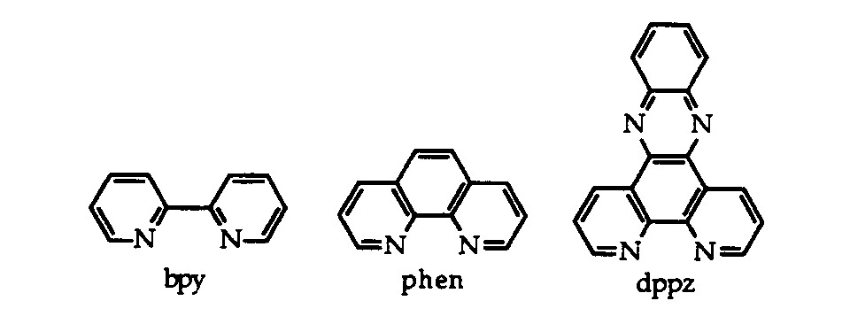
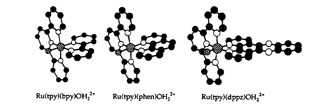
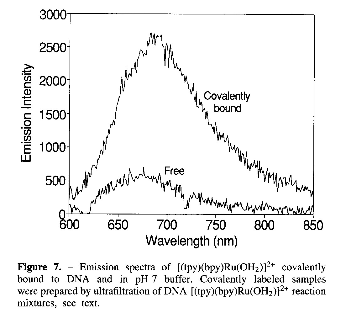
I began my training as a chemist at North Carolina State University.
My research experience as an undergraduate concerned the interaction of
small metal complexes with DNA. In that work, we used the characteristic
optical properties of transition metal complexes to study the mechanism
of binding and strength of interaction of small ruthenium coordination
complexes with nucleic acids. The binding of such complexes has been
used to determine structure of nucleic acids and in some cases to create
new therapies for disease. My involvement in this research convinced
me that I should attend graduate school and study bioinorganic chemistry.
|
|
I completed my PhD training in 1997 at the University of North Carolina,
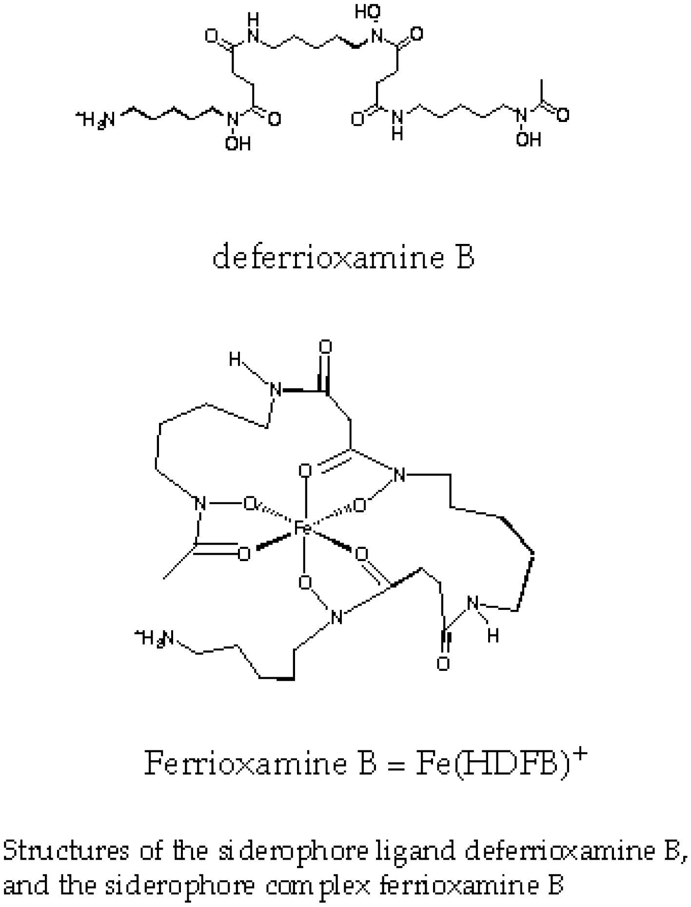
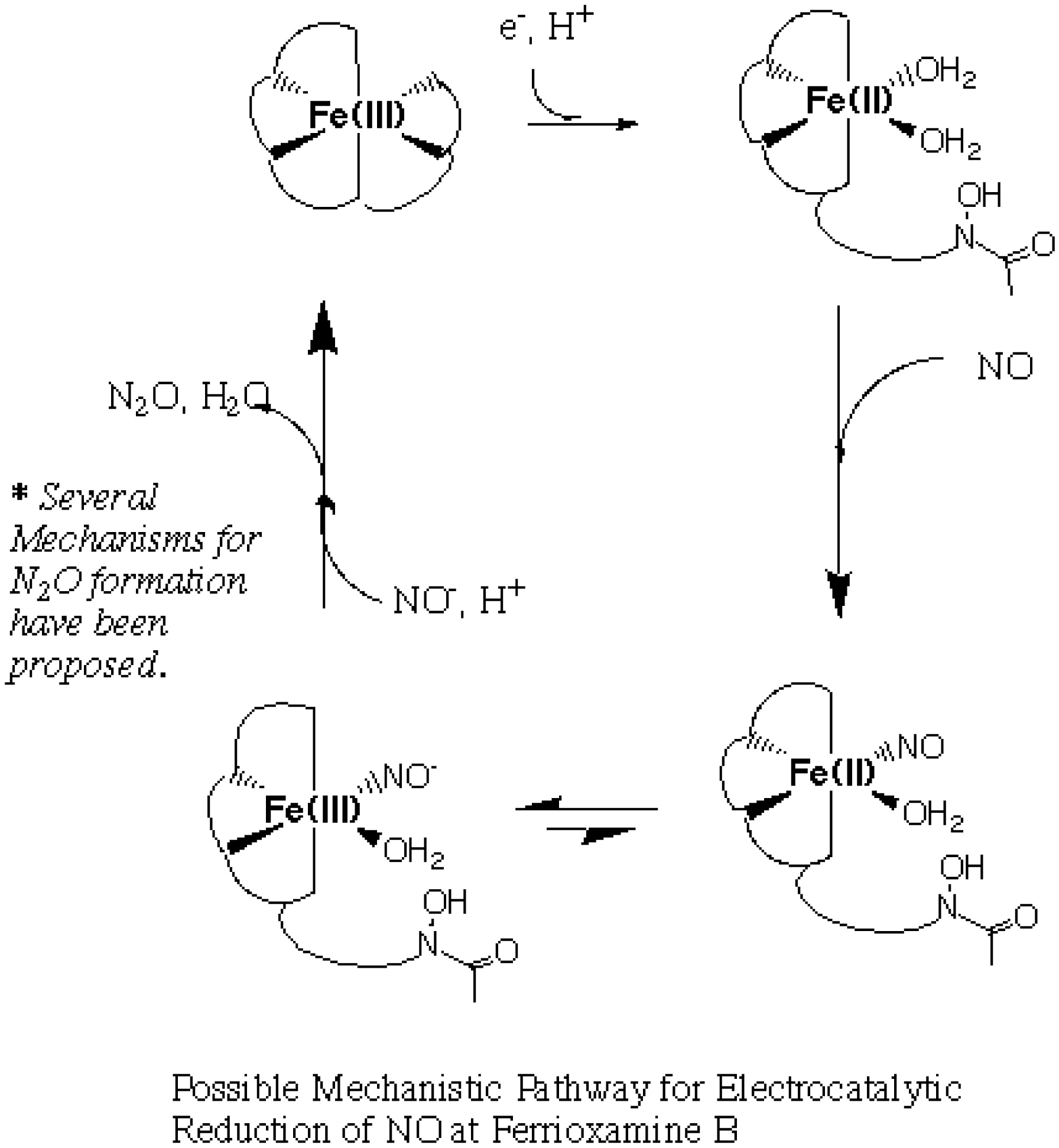
under the guidance of Professor Holden Thorp. My doctoral research
focused on the reduction of small molecule substrates like nitric oxide
catalyzed by an iron-containing class of biomolecules called siderophores.
This work has fascinating implications in biological and environmental
systems. NO has long been recognized as a pollutant, and much of
the focus on NO reseach has been aimed toward developing systems which
can mediate the contribution of NO to environmental problems. The discovery
in recent years of NO's role as a biological signalling agent has shifted
the focus somewhat towards the mechanisms of production, regulation and
recognition of NO in biological systems. My graduate research fell
at a point somewhere in between these two fields of NO research.
The discovery that ferrioxamine B, a biologically-derived iron complex,
mediated mortality caused by excess NO production during septic shock,
led to an exploration of the suitability of complexes similar to ferrioxamine
B as reduction catalysts for NOx compounds. This
project will be ongoing in my lab at UMD.
|
|
My first post-doctoral position involved the determination of the structure
and role of the iron sulfur cluster(s) in an anaerobic metalloenzyme called
pyruvate-formate lyase activase. This enzyme plays a key role in
the ability of certain bacteria to survive in the absence of oxygen.
Following anaerobic overexpression and purification of the enzyme from
e. coli, the metal centers were studied by biochemical assay and spectroscopic
methods.
|
|
My second post-doctoral position has focused again on the structure
and role of metals in metalloenzymes, particularly focusing on the magnetic
properties resulting from the presence of unpaired electrons on the metal.
|
|
The point of all this is to say that the study of metals in biological systems encompasses a broad range of problems and the presence of metal ions opens up an extensive array of biophysical techniques for use in the study of the structure and role of these metals.
 1992-1997